Contributing physicians in this story
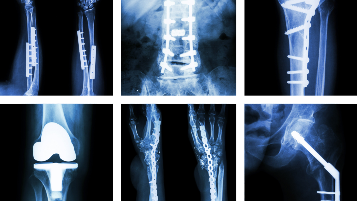
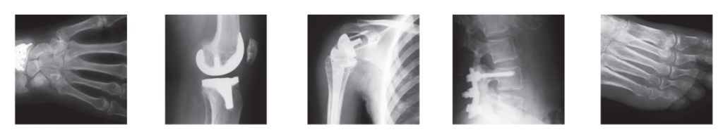
If you have a bone or musculoskeletal problem, your physician will usually begin the diagnostic process with x-rays. X-rays are an excellent screening tool for visualizing bone pathologies, such as fractures or degenerative joint disease. In many instances, however, either the presence or absence of findings will warrant a closer look, prompting your attending physician to order a computed tomography (CT), magnetic resonance imaging (MRI), or nuclear medicine scan. Each of these scans offers different advantages and disadvantages for you as a patient, and the type of imaging study or studies your physician orders will depend largely on your symptoms and health history.
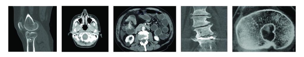
Computed tomography
The word tomography comes from the ancient Greek roots, tom-, meaning a cutting or slice, and –graphy, meaning imaging or writing, and thus refers to the imaging of internal cross-sections of the body. The term computed tomography (CT) or, sometimes, computed axial tomography (CAT), refers specifically to combined computer-processed x-ray images of internal cross-sections of the body taken from different angles. If you suffer an acute trauma, your doctor will typically order a CT scan to rule out fractures and dislocations. Although a CT scan may be recommended for you largely because of its ability to evaluate bony structures, special studies, such as a CT arthrography (joint imaging) or CT myelography (spinal cord, nerve root, and other soft tissue imaging), may be ordered to provide details about your soft tissue. By injecting a radiocontrast agent or medium (typically an iodine or barium compound) directly into the joint or spinal canal to make internal structures and bodily fluids more visible on imaging, such studies can offer detailed pictures.
One advantage of a CT scan over other types of scans is how quickly it can be performed. During a CT scan, a high-powered x-ray tube rotates around your body to generate the images. This generally takes only a few minutes and can be performed with a single breath hold. Another advantage of CT imaging is that it is safe even if you have had a metal device or certain types of hardware implanted in your body. In fact, if this is the case for you, your doctor should order a CT scan rather than other imaging modalities.
The major disadvantage of having a CT scan is that it uses ionizing radiation (the emission of electrical charges and electromagnetic waves) to produce images and in the process exposes you to a higher dose of radiation than a conventional x-ray. Radiation exposure can harm cellular structures and processes and can directly or indirectly affect DNA. However, as CT scans are now performed more quickly, the amount of radiation to which you are exposed is less than you would receive as an airline passenger on a long flight. Furthermore, advances in CT scanning have resulted in higher resolution images and increased comfort for you as a patient.
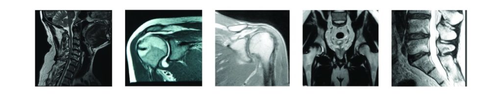
Magnetic resonance imaging
Unlike CT scans, which use x-rays, MRI scans use powerful magnetic fields along with radio frequency pulses to produce detailed images of bones as well as soft tissues such as muscles, tendons, ligaments, and cartilage. Since the images are produced by a magnetic field, you are not exposed to radiation, and this constitutes an advantage over a CT scan.
Additionally, MRI scans are superior for soft tissue imaging. As with soft tissue CT scans, administering contrast agents or mediums (usually a compound with the ability to alter the magnetic properties of nearby hydrogen atoms) through an IV has been found to enhance imaging. Moreover, the contrast setting of an MRI can be changed to highlight different types of tissues and any subtle differences between them. The imaging plane can also be changed without moving the patient.
The chief disadvantage of an MRI is that its powerful magnetic field can prove harmful if you have certain metal devices in your body such as pacemakers, cardiac monitors, spinal cord stimulators, and some types of surgical clips. Another disadvantage of this type of imaging is the time required to scan—typically around 30 minutes. Additionally, when you have an MRI, you are placed in a cylindrical tube that could make you feel uncomfortable or claustrophobic. Open MRI scanners are available, but their magnetic field is significantly weaker, and, consequently, they produce much less detailed images. Overall, despite their disadvantages, including the increased cost compared with CT scans, MRIs are still more commonly performed when a high degree of soft tissue detail is needed.
![]()
Nuclear medicine
Nuclear medicine refers to a type of noninvasive, painless (apart from the placement of an IV) imaging procedure that helps physicians diagnose and evaluate a host of medical conditions involving bones or soft tissue. As a diagnostic tool, nuclear medicine has been around for 60 years—longer than CT or MRI. In contrast to both CT and MRI scans, nuclear medicine imaging uses radioactive materials called radiopharmaceuticals or radiotracers. These drugs, which can be taken either orally or intravenously, contain radioactive isotopes (variants of elements that emit energy in the form of gamma rays and decay quickly). Consequently, with nuclear imaging, unlike x-rays and CT scans, the radiation that helps produce the images is emitted by your body rather than externally generated. This radiation can then be detected by a specialized camera or imaging device. From the data gathered, the device produces scintigrams (recordings of the scintillation or flashing from the radioactivity) that yield images of your bones. Areas that take up more or less of the radiopharmaceutical show up as either brighter or darker than normal on a scintigram and may indicate abnormalities in your bone.
The primary drawback of nuclear medicine imaging is that it exposes you to ionizing radiation. You could also have an allergic or injection site reaction to the radioactive material. On the other hand, nuclear medicine imaging offers a huge advantage over other types of scans: rather than merely picturing structures, nuclear scans can show how different areas or organs of the body are functioning on the molecular level. This means they have the potential to detect disease in its earliest stages. For instance, a bone scan can often pick up abnormalities inside your bone that may indicate cancer much earlier than a normal x-ray. It can also reveal a tiny fracture before it can be seen on an x-ray.
Understanding your test
If you go to your doctor with a bone or musculoskeletal problem—for example, a stress fracture, torn tendon, or even complications from a hip or knee replacement—he or she may order a CT, MRI, or nuclear scan. The proper imaging modality will depend on your particular situation. Thus you should feel free to discuss any questions or concerns you may have about the diagnostic process or a particular test with the ordering physician or a radiologist (an expert at reading such images). You may also want to gather information on your own ahead of time. A website dedicated exclusively to MRI safety can be found at www.MRIsafety.com. Informing yourself about different types of diagnostic imaging is important so that you can feel confident that your doctor is prescribing the most appropriate imaging modalities for your condition and that the testing you have performed is safe.
Author: Cameron C. Kersey, MD | Columbus, Georgia
Volume 28, Number 2, Spring 2016.
Last edited on October 18, 2021